Best medical annotation tools for Healthcare AI
About the series
Are you unsure which annotation tools are best suited for your healthcare project?
At Humans in the Loop, we offer annotation services for medical data such as X-rays, MRI and CT scans, microscopic imagery, ultrasound videos, and other 2D, 3D, and video data. Our global workforce of medical annotators comes from conflict-affected and displaced communities in various locations worldwide.
We are pleased to share our experts insights on the advantages and disadvantages of the top medical annotation tools.
1. 3D Slicer

The 3D Slicer segmentation tool is one of the most powerful and versatile tools available to medical professionals today. This open-source software is used to delineate regions in an image, typically corresponding to anatomical structures, lesions, and other object spaces.
The 3D Slicer segmentation tool is an essential tool for medical research and clinical practice, thanks to its wide range of features. One of its key features is its ability to perform precise segmentation, which accurately identifies each structure or region of interest. This feature has made it invaluable for medical professionals in radiology, oncology, and neurology who need to extract specific information from medical images.
In addition to its segmentation capabilities, 3D Slicer has many uses in the medical field:
- DICOM standard interoperability “Support of a wide range of DICOM information objects, such as 2D, 3D, 4D images”.
- Image segmentation. “Lung CT segmentation and analysis for COVID-19 assessment”
- Artificial Intelligence.
- 3D Modeling and 3d Printing.
- Surgical planning and guidance.
A key advantage of the tool is its integration with other medical imaging tools. This integration enables users to directly import and export medical images and segmentation files from other software applications, such as MRI and CT scan machines. However as an offline tool, it lacks quality control and management features that would allow you to track performance and easily manage all of your project’s tasks.
Our opinion :
We recommend it for 3D segmentation of CT scans as it has many modules which allow you to deal with different types of CT scans. The Slicer RT module is one of them that allows you to read and write DICOM Radiation Therapy objects (RT structure set, dose, image, plan, etc.) and provides tools for visualizing and analyzing them.
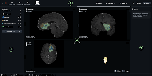
2. Encord

Encord is a medical annotation platform that utilizes computer vision to accurately and efficiently classify and segment medical images. It is a collaborative DICOM & NIfTI annotation platform for computer vision graduates from ITK Snap & 3D Slicer, integrated with Encord’s collaborative annotation platform.
The Encord platform offers many benefits to healthcare providers and researchers.
First, it allows for more accurate and consistent annotations of medical images, leading to better diagnoses and treatments. Encord supports a range of file formats, including DICOM and NIfTI. Its DICOM tool has been developed alongside clinicians and healthcare data scientists, delivering efficient functionality and a seamless user experience when annotating specialized datasets. This ensures maximum efficiency and usability for end-users.
Additionally, Encord is the only computer vision data platform that supports labeling videos of any length. This means any video can be uploaded, regardless of its format or length, ensuring your model has the best datasets for detecting cancerous polyps, ulcers, IBS, or any other condition. Medical professionals can quickly annotate image data without sacrificing accuracy. This can be especially helpful in situations where rapid diagnoses are required, such as emergency rooms or critical care settings.
Gastroenterology Segmentation and Annotation” Colonoscopy video annotation features:
- Track polyps in the video
- Colonography and scope videos in one tool
- Support Mayo and UCEIS scoring rubrics
Our opinion :
Encord is an incredibly valuable tool for healthcare providers and researchers who rely on medical imaging to diagnose and treat various conditions. The platform’s ability to accurately and efficiently annotate image data, adapt to new information, and enhance diagnostic accuracy make it an essential tool in modern healthcare.
3. OHIF
OHIF (Open Health Imaging Foundation) is a nonprofit organization that develops open-source software frameworks for medical image viewing, processing, and analysis. The OHIF annotation tool is a web-based application designed to facilitate the annotation of radiological images by radiologists and clinical researchers.
The OHIF annotation tool is built on top of the OHIF viewer, which is an open-source tool for loading and viewing various medical imaging formats. The viewer can retrieve and load images from most sources and formats, render sets in 2D, 3D, and reconstructed representations, and enable the manipulation, annotation, and serialization of observations. Additionally, it supports internationalization, OpenID Connect, offline use, hotkeys, and many other features. All of the viewer’s core features are built using its own extension system, which provides extensibility and enables us to offer:
- 2D and 3D medical image viewing
- Multiplanar Reconstruction (MPR)
- Maximum Intensity Project (MIP)
- Whole slide microscopy viewing
- PDF and Dicom Structured Report rendering
- User Access Control (UAC)
- Context-specific toolbar and side panel content
The annotation tool also provides a range of customization options for users. Users can modify the color, size and shape of annotations, as well as add labels and notes to provide context and additional information about the annotations.
Our opinion :
The OHIF annotation tool is a powerful and flexible tool that facilitates collaboration between radiologists and clinical researchers. Users can share annotations and other imaging data with colleagues. Additionally, it can be integrated with other third-party software applications, such as machine learning algorithms, to assist radiologists in the diagnosis process. Moreover, the OHIF annotation tool is designed to be extensible and customizable.
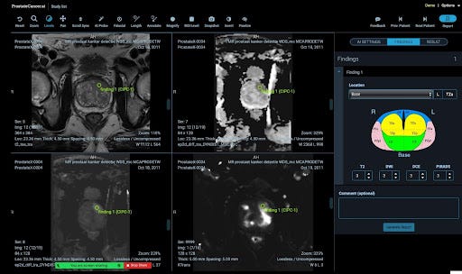
4. RedBrick
RedBrick is an innovative platform that enables medical practitioners to securely and efficiently annotate and share medical data. It was launched in November 2021 and created by Shivam Sharma and Derek Lukacs.
RedBrick is a purpose-built SaaS application designed to help healthcare AI teams annotate medical data more effectively. It offers annotation tools for CTs, MRIs, X-rays, etc., as well as comprehensive project management and quality control tools.
RedBrick AI also provides a web-based DICOM annotation tool with native support for DICOM medical images. It supports 2D and 3D data and allows for segmentation, classification, and vector annotations. The platform provides an intuitive and user-friendly interface, designed to be easy to use, even for those with limited technical experience. This ensures that medical professionals can spend less time navigating complex software.
Segmentation tool features :
- Brush
- Pen
- Region Growing
- Contour Tool
- Creating a contour & interpolation
- Editing a contour
- Smart contouring
- Island Removal
- Hole Filling
- Paint Bucket
- Thresholding
Our opinion :
The Redrick medical annotation platform is a game-changer in the world of medical annotation. Its innovative features provide medical professionals with a comprehensive solution for annotation and analyzing medical data securely and efficiently as it provides specialized annotation tools accessible via a web browser and integrated into your existing data storage system. With its medical data annotation tools for imagery, it aims to lay the groundwork for healthcare artificial intelligence.
5. Supervisely
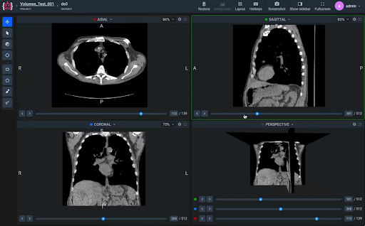
Supervisely medical interface was released in August 2017 and provides a well-known intuitive interface to view and manipulate volumetric medical images in multiple projections and slices with expert features for CT and MRI in 2D or 3D. This software tool has helped to revolutionize the process of medical imaging data annotation, making it quicker, more efficient, and with fewer errors.
The Supervisely medical interface has made this process more effective and efficient, by providing a simple, user-friendly, and interactive interface that allows medical professionals to annotate medical images quickly and accurately.
Interface features:
Navigation through slices in multiple projections
Perspective 3D view panel
Multi-window layout
Windowing, windowing presets
Crosshair tool
Rotate, flip. invert colors
Colormaps
Multi-planar labeling
Features:
Multi-slice annotation objects
Building 3D volumes from 2D figures
Our opinion : We recommend it for beginners and simple projects as it provides an easily operated user interface and multi-planner labeling for DICOM annotation as it provides a variety of features that enable healthcare professionals to ensure that their medical image annotations are accurate. These features include the ability to adjust the image brightness and contrast, zoom in and out, and rotate the image for a better view. This software tool also allows healthcare professionals to create multiple labels and tags for each image.
6. V7 Labs

The human visual Cortex consists of 6 regions”V1 to V6″ Each region has a specific function in recognizing shapes colors, forms, and motion. In 2018, Alberto Rizzoli and Simon Edwards Founded V7 and wanted the V7 to bring a similar revolution — granting all automata with sight, and enabling them to multiply the tasks they can perform.
This innovative platform is designed to simplify complex medical documentation, saving time and increasing accuracy in the process.it is FDA and HIPAA-compliant. It can be used in many medical specialties including Radiology, Dermatology, Pathology, and Dentistry. Also, it supports the DICOM format which is implemented in almost every radiology, cardiology imaging, and radiotherapy device (X-ray, CT, MRI, ultrasound, etc.), and increasingly in devices in other medical domains such as ophthalmology and dentistry. With hundreds of thousands of medical imaging devices in use.
In addition, V7 provides an auto-annotation for 3D medical images that speeds up your annotation time by using AI models to annotate your data. This feature is suitable for easy projects and high-quality datasets but won’t add value in complicated projects as it can’t provide an accurate annotation and will consume a lot of time to adjust the AI Model annotation so we don’t recommend it in medium or high difficulty projects.
Advantages of V7’s DICOM annotation features:
- orthogonal views
- image manipulation
- windowing
- consensus stage
Our opinion :
V7 is a powerful annotation tool. Its intuitive and user-friendly design, powerful annotation tools, and commitment to data security and privacy make it an ideal solution for healthcare professionals who are looking to optimize their workflow and improve patient outcomes.
Hope this was helpful! If you are working on an AI project and are currently reviewing which tool might be the most appropriate for it, get in touch with us and we would be happy to have a call and advise you on the best way to build your pipeline.
